Authors: William L. Rice, Alfred N. Van Hoek, Teodor G. Păunescu, Chuong Huynh, Bernhard Goetze, Bipin Singh, Larry Scipioni, Lewis A. Stern, Dennis Brown
DOI: 10.1371/journal.pone.0057051
Abstract Summary
Helium ion microscopy achieves breakthrough sub-nanometer imaging of rat kidney tissue, revealing unprecedented details like pores in glomerular filtration slits and delicate membrane structures on collecting duct cells. This novel technique preserves tissue architecture better than conventional electron microscopy and enables multi-target labeling with gold probes, opening new possibilities for studying cell surface ultrastructure.
Why Brain? 🧠
Novel helium ion microscopy achieves sub-nanometer resolution imaging of rat kidney tissue, revealing unprecedented detail of cellular structures like podocyte membranes and filtration pores.
License: CC BY.
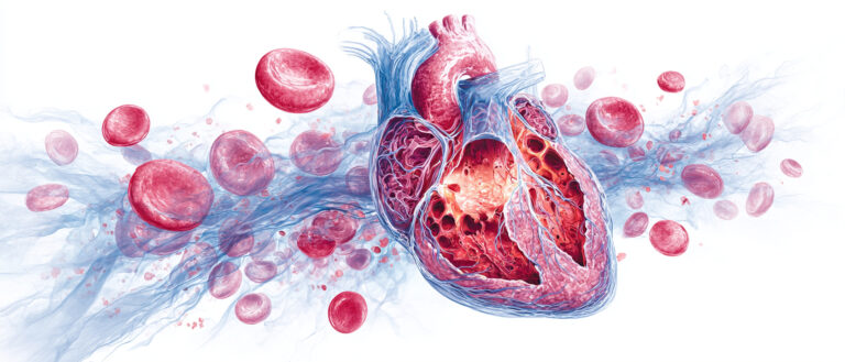
The image is AI-generated for illustrative purposes only. Courtesy of Midjourney.


